CT Scans of Ancient Egyptian Mummies Reveal Lifespans, Health, and Daily Life of Priests
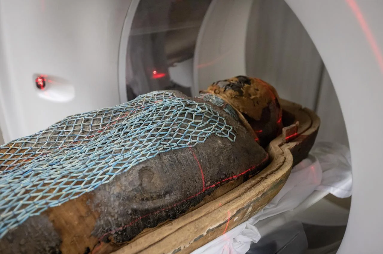
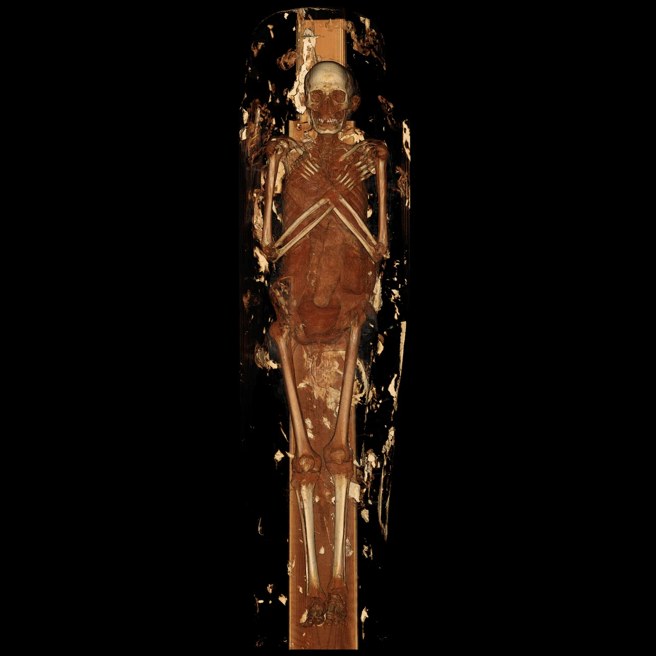
Radiologists at Keck Medicine of USC have examined two ancient Egyptian mummies using high-resolution CT scanning, producing exceptionally detailed views of bodies preserved for more than two millennia. The study focused on two priests—Nes Min, who lived around 330 BCE, and Nes Hor, who lived around 190 BCE. Each mummy was scanned while still resting in the lower half of a massive sarcophagus weighing roughly 200 pounds.
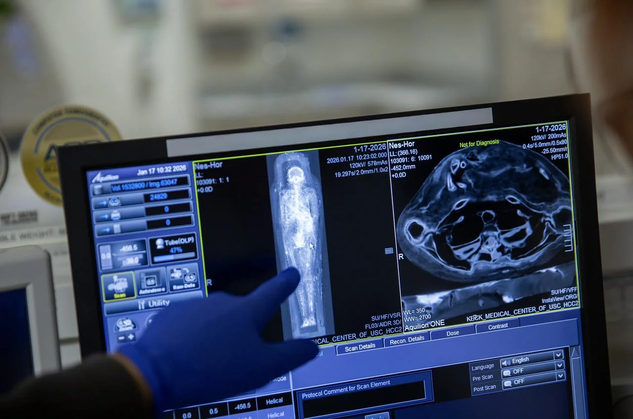
The scanner captured hundreds of thin cross-sectional images across each body, which specialists then assembled into three-dimensional digital models. These reconstructions revealed remarkably fine physical details, including eyelids, lips, and facial bone structure, offering a clearer sense of each individual’s appearance beyond the linen wrappings.
The scans also documented signs of aging and disease. Nes Min showed significant damage in the lower spine, including a collapsed lumbar vertebra—an injury pattern associated with long-term strain and age-related degeneration similar to chronic back conditions seen in modern patients. Nes Hor exhibited a different health profile, with advanced tooth decay and severe deterioration in one hip joint. Such joint damage likely caused pain and restricted movement, and the overall bone condition suggests that Nes Hor lived to an older age than Nes Min.
CT imaging revealed burial objects hidden within the wrappings as well. Nes Min was interred with small items shaped like scarab beetles and a fish, artifacts that had remained unseen for more than 2,000 years. The scans allowed researchers to measure and study these objects without physically disturbing the mummies.
Following the imaging work, visualization specialists created digital reconstructions of the skeletons and selected artifacts, then produced life-size 3D prints of skulls, spines, hips, and burial items using medical-grade printers. These replicas will be displayed alongside the mummies and interactive digital content at an exhibition at the California Science Center, opening February 7 as part of Mummies of the World.
The same imaging and 3D-printing workflow underpins modern clinical care. Hospitals routinely use CT and MRI data to build digital models of organs such as the heart, liver, or pelvis, enabling surgeons to plan complex procedures, choose implant sizes, and rehearse difficult steps. Nearly two dozen printers at the USC Center for Innovation in Medical Visualization support both patient care and research projects like the mummy study.
Physicians note that holding a physical model also improves communication with patients, making anatomy and planned procedures easier to understand. The mummy project demonstrates how medical imaging technologies designed for living patients can also illuminate the lives of ancient people—preserving fragile remains while revealing health, injury, and aspects of daily life in ancient Egypt.