Pediatric Neurosurgery, 3D Printing, and Life: The Miraculous Return of Bentley Yoder After Encephalocele
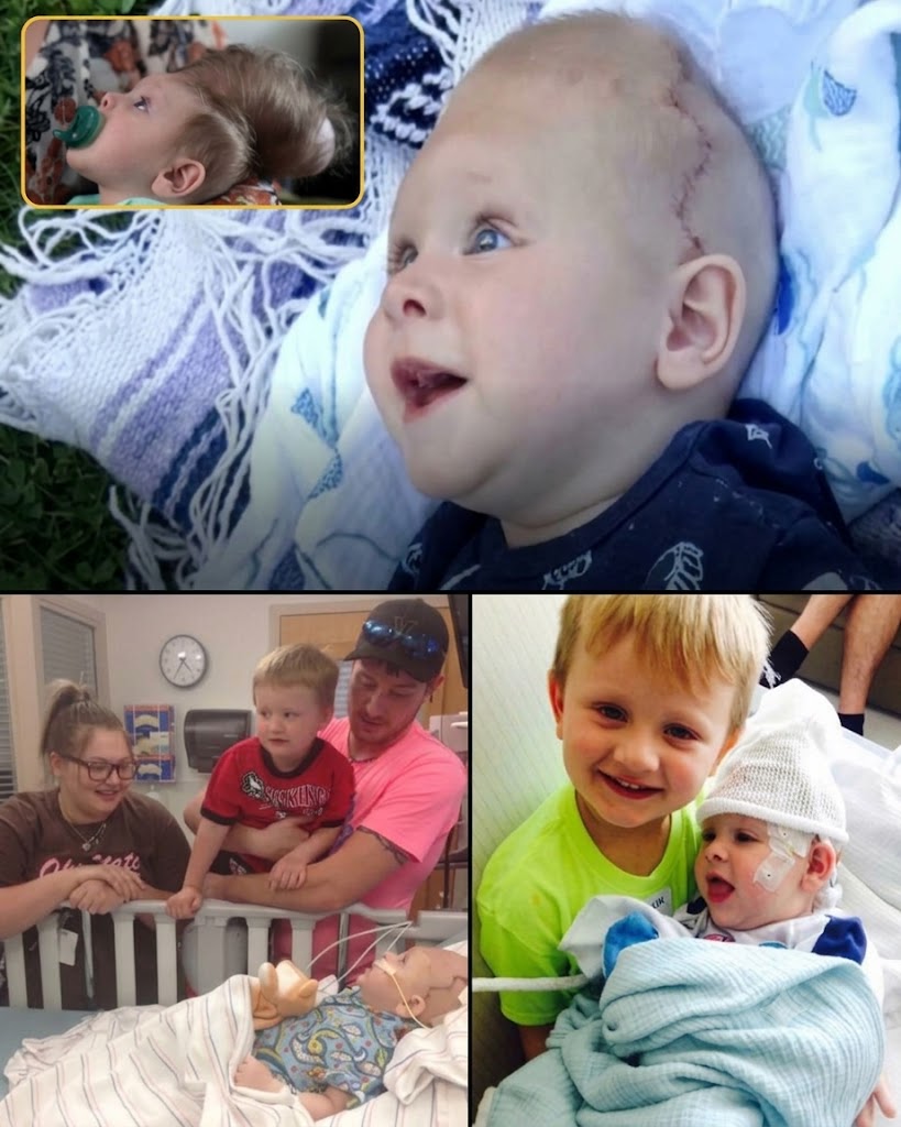
At 22 weeks of pregnancy, a routine ultrasound in Ohio delivered news no parent expects. Sierra and Dustin Yoder learned their unborn son had a large occipital encephalocele: a significant portion of his brain was developing outside his skull. Doctors described the condition as incompatible with life. They predicted that if the baby survived birth at all, he would have no meaningful cognition, speech, or movement. Termination was strongly recommended.
The couple scheduled the procedure, then changed their minds the night before. They chose to continue the pregnancy and give their son whatever time he had, even if it was only hours of palliative care.
Bentley Ross Yoder was born on November 1, 2015, after a normal vaginal delivery. He cried vigorously, breathed on his own, and surprised every clinician in the room. In the weeks that followed he continued to defy predictions: he fed, responded to his parents and older brother Beau, and showed no seizures. The sac on the back of his head, however, grew rapidly as it filled with cerebrospinal fluid. The thin covering of skin stretched over functional brain tissue.
Local neurosurgeons proposed simply amputating the mass, assuming the extruded tissue was damaged and unused. Sierra and Dustin believed otherwise. Their son’s movements and alertness suggested he was using that tissue. A second opinion at the Cleveland Clinic confirmed their suspicion. Neurosurgeon Violette Recinos referred them to Boston Children’s Hospital, whose craniofacial and neurosurgical teams treat some of the most complex encephalocele cases in North America.
There, plastic surgeon John G. Meara and neurosurgeon Mark R. Proctor examined Bentley. Imaging showed that the protruding brain included regions controlling vision, motor function, and problem-solving—roughly 100 cubic centimeters of living tissue that could not be discarded. The only viable option was to expand the undersized skull and gently return the brain to the cranial vault.
The team turned to Boston Children’s newly expanded surgical simulation center. High-resolution scans were converted into physical 3D-printed models of Bentley’s skull and encephalocele. Surgeons practiced osteotomies, measured how much additional volume they could create, and refined the sequence of steps before ever entering the operating room. The models allowed them to decide which small sliver of tissue was non-viable and could be left out, and which portions had to be preserved.
On May 24, 2016, when Bentley was nearly seven months old—old enough for his skull to be structurally sound, yet before the sac risked rupture—the operation took place. Surgeons drained excess cerebrospinal fluid, made controlled cuts in the calvarium, inserted bio-absorbable plates to hold the expanded bone in position, and eased the brain back inside. Leftover bone fragments helped close the defect. The procedure lasted several hours and achieved its primary goals. Two later operations placed a shunt to manage hydrocephalus.
Bentley recovered. He retained vision in both eyes. Within weeks he was smiling, babbling, holding his head up for longer periods, and interacting like other infants his age. Doctors cautioned that his long-term developmental trajectory remained unknown; encephalocele often brings delays, and his uniquely rearranged brain had no exact precedent. Yet the fact that he was meeting early milestones was itself a gift his parents had been told they would never receive.
The case illustrated how 3D printing and multidisciplinary simulation can turn an operation once considered impossible into a planned, rehearsed procedure. It also illustrated the limits of prenatal prognosis. Bentley’s parents were told there was “zero chance.” He left Boston Children’s Hospital alive, seeing, and smiling.
Years later the family continued therapies closer to home in Ohio. Public updates after 2017 are scarce, as is common once the acute medical crisis recedes. What remains documented is a child who was not supposed to live past his first hours, yet whose brain was returned to his skull through careful planning, advanced imaging, and a refusal to accept the first diagnosis as the final word.
Sources
- Boston Children’s Hospital video series “Bentley’s Second Chance” (2016)
- STAT News, “Daring brain surgery gives baby with a birth defect a chance,” June 15, 2016
- The Washington Post, “‘We had no hope’: The amazing story of a baby born with his brain outside his skull,” June 20, 2016
- Vector / Boston Children’s Hospital, “Printing your skull: How 3D printing helps surgeons plan complex encephalocele brain surgeries,” July 2016
- ABC News, “Baby Thriving Despite Condition Thought Incompatible With Life,” June 2016
- 3DPrint.com and contemporaneous reporting from Boston Magazine, Gizmodo, and The Times-Reporter (Sugarcreek, Ohio), 2016–2017