🚨 NEUROSURGERY, FAITH, AND LIFE: THE MIRACULOUS PROCEDURE THAT SEPARATED SISTERS JOINED AT THE SKULL
Sisters Joined at the Skull Separated After Staged Surgery Planned in 3D
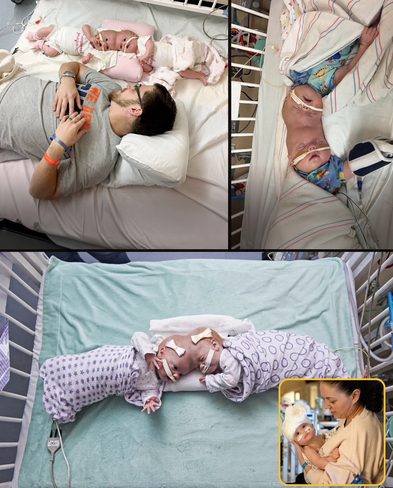
Craniopagus twins — infants fused at the skull, often sharing veins and sometimes brain tissue — are among the rarest surgical challenges in pediatric neurosurgery. When the girls face opposite ways, they may never have seen each other’s faces until the last connections are cut.
Two-year-old sisters Safa and Marwa Ullah, born in Pakistan and treated at Great Ormond Street Hospital in London in 2018–19, match that pattern. Joined crown to crown, they shared bone and a complex venous system. A team led by neurosurgeon Owase Jeelani and plastic surgeon David Dunaway spent months on virtual-reality models and 3D-printed anatomy before three operations totaling about 50 hours. One stage ran more than 20 hours. After the final division they could be placed on separate beds and, for the first time, look at one another.
Other landmark cases use the same toolkit. In 2002 at UCLA, Guatemalan sisters Maria de Jesus and Maria Teresa were separated in a roughly 20- to 23-hour operation by a team of about 50. A nurse held a mirror so one child could see her sister. In 2016 at Montefiore, Jadon and Anias McDonald (boys) were divided in a 27-hour case built on 3D prints. In 2020 in Rome, Ervina and Prefina, posterior craniopagus sisters, were separated in an 18-hour final procedure after staged work at Bambino Gesù.
Success is measured in preserved vision, movement and independent skulls — not only in the moment two children first meet face to face. Every published series stresses that outcomes still vary and that the work is staged, not a single unbroken miracle shift.
Source: Great Ormond Street / BBC reporting on Safa and Marwa; UCLA Health on Maria de Jesus and Maria Teresa (2002); contemporaneous accounts of Montefiore and Bambino Gesù craniopagus separations.