Vincent Oketch and the Legs That Outweighed the Child
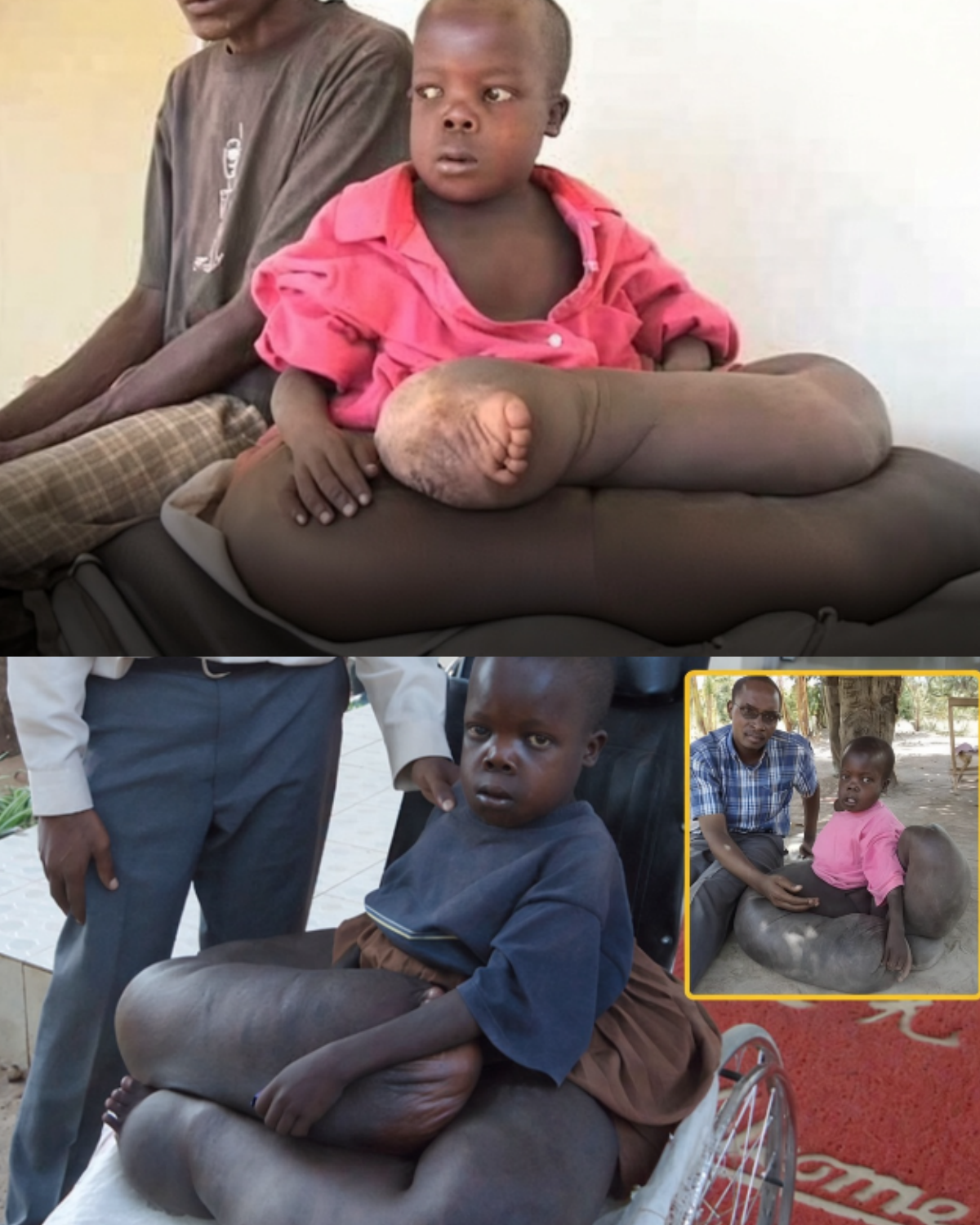
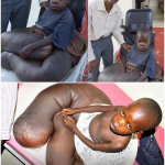
Vincent Oketch lived in Peta, a village in Uganda. Swelling in his legs began around 18 months of age. His parents, Tito Opoya among them, were too poor for a clinic. By ten, in 2012, the limbs were so large he could barely crawl. Shorts would not fit; he wore dresses and was carried on a blanket. Other children teased him.
Dr Isaac Osire, who ran a small charity (EDYAC) for village children, drove him to Kampala. MRI and biopsies produced a stack of labels: failed lymphatics, elephantiasis, tissue deformity, a dislocated hip, broken ankles—and a suggestion of necrotising fasciitis. Hospital staff discussed amputation at the thigh. Osire asked publicly for a British or other foreign specialist because the diagnoses contradicted one another.
The photographs in the collage—the pink shirt on a bench, the wheelchair, the man in a checked shirt with a hand on the swollen limb—are the Barcroft / Mirror pictures from that appeal.
Lymphatic filariasis (worm-borne “elephantiasis”) and podoconiosis (non-filarial swelling from volcanic soil) both exist in East Africa. A congenital lymphatic malformation can look the same. Necrotising infection on top of any of those can kill. In 2012 the Kampala team did not publish a clean, single diagnosis or a follow-up of Vincent walking. Later viral videos recycle the same stills and invent a finished “international rescue.” That ending is not in the contemporaneous record.
What is documented is a ten-year-old whose legs weighed more than the rest of him, a village without money, and doctors who were honest about not knowing which knife, if any, to use. Stigma and “curse” talk are real in such cases. So is the limit of a newspaper appeal: it can move a child to a city hospital. It does not automatically drain, reconstruct, or restore a gait.
Sources
- Daily Mail / The Mirror, 15 May 2012: Vincent Oketch, Peta, Uganda; Dr Isaac Osire / EDYAC; Kampala imaging and amputation discussion
- News Ghana / contemporaneous wires: mixed diagnosis (lymphatic failure, possible filariasis, possible necrotising fasciitis)
- Regional context: lymphatic filariasis and podoconiosis in Uganda