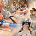
Manar Maged: The Extraordinary Story of an Egyptian Baby Born With a Parasitic Twin

In 2004, the birth of an Egyptian baby named Manar Maged drew international attention because of one of the rarest congenital conditions ever documented: craniopagus parasiticus. The condition occurs when a developing embryo begins to form identical twins but the division is incomplete, leaving a severely underdeveloped parasitic twin attached to the more fully developed twin. In Manar’s case, the parasitic twin consisted essentially of a second head attached to her skull. Medical literature describes craniopagus parasiticus as extraordinarily rare, with estimates of only a handful of documented cases.
Manar was born on March 30, 2004, in Egypt. The second head was attached to her own and was incapable of independent survival because it did not have a functioning body. Nevertheless, doctors reported that the attached head could blink and make facial movements, including smiling. The unusual condition presented an enormous medical challenge because the two structures shared important blood vessels and were closely connected around the skull.
Doctors at Benha Children’s Hospital, north of Cairo, spent months evaluating Manar before attempting separation. At approximately 10 months old, she underwent a highly complex operation in February 2005. Contemporary reports from Reuters described the procedure as lasting about 13 hours, with surgeons carefully separating the parasitic head and controlling the blood supply between the two structures.
The operation was considered a major achievement. Immediately afterward, doctors reported that Manar was breathing normally, had a normal heartbeat and blood pressure, could move all four limbs and showed no signs of paralysis. She remained in intensive care while physicians closely monitored her recovery.
However, surviving the separation was only the beginning of Manar’s medical struggle. Within weeks, she developed hydrocephalus, a condition in which excess cerebrospinal fluid accumulates inside the brain. In March 2005, doctors performed another operation to implant a valve designed to drain the excess fluid. Reports said the procedure helped control the seizures associated with the condition.
Manar subsequently faced additional complications and recurrent infections. Despite the initial success of the separation, her health remained fragile. In March 2006, she was admitted to hospital with a severe infection. Doctors reported that the infection had affected her brain and that her condition deteriorated rapidly. She died on March 25, 2006, shortly before her second birthday.
Manar’s case remains significant in the history of pediatric neurosurgery because it demonstrated both the possibilities and the limitations of modern medicine when confronted with extraordinarily rare developmental abnormalities. A later neurosurgical publication discussing her case noted that she had survived the separation without neurological paralysis for many months before eventually dying from a severe brain infection.
Craniopagus parasiticus itself remains poorly understood. Researchers believe it is related to incomplete separation during the development of identical twins, but because the condition is so rare, the precise biological mechanisms remain uncertain. Medical reports have documented only a very small number of cases, and survival has historically been exceptional.
Manar Maged’s story is therefore more than an unusual medical case. It represents the extraordinary risks faced by children born with rare congenital conditions and the determination of doctors and families to pursue treatment even when the chances of long-term survival are uncertain. Her 2005 operation pushed the boundaries of pediatric surgery at the time, while her later complications also demonstrated how difficult recovery can remain even after an apparently successful procedure.
Although Manar’s life was tragically short, her case became part of medical history. It continues to be referenced in discussions of craniopagus parasiticus and the challenges involved in separating conjoined structures involving the skull and brain. Her story remains a poignant reminder of both the remarkable capabilities of modern medicine and the limits it can encounter when confronting conditions that occur only a few times in millions of births.
Sources
- Journal of Medical Case Reports / PubMed — “Craniopagus parasiticus – a parasitic head protruding from temporal area of cranium: a case report.” Provides medical background on the condition, its rarity and previous documented cases, including Manar Maged.
- Reuters via Gulf News — “Baby stable after second head removed, say doctors.” Contemporary report on Manar’s 13-hour separation surgery and her immediate postoperative condition.
- Reuters via Al Jazeera — “Two-headed baby has second surgery.” Contemporary report on Manar’s postoperative hydrocephalus and treatment with a brain valve.
- ABC News / Reuters — “Toddler with two heads dies.” Report on Manar’s death in March 2006 following a severe brain infection.
- PubMed — “Successful separation of craniopagus parasiticus.” Neurosurgical report describing the technical challenges of separating Manar’s parasitic twin and the medical significance of the procedure.