What the Photographs Show—and What They Do Not: Extreme Childhood Hydrocephalus
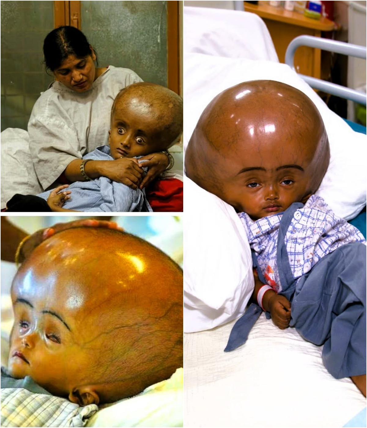
The images show a toddler whose skull has been stretched by severe hydrocephalus: a globe-like head, taut shiny scalp, prominent veins, and eyes that appear sunken or forced downward. The accompanying caption identifies the child only as three years old. No verified name, hospital, cause, operation, or later outcome is attached to these particular frames. That gap is the most important fact about the pictures.
Hydrocephalus is the buildup of cerebrospinal fluid inside the brain’s ventricles. In infants and young children the cranial sutures have not fused, so prolonged pressure can enlarge the head far beyond normal. Causes include congenital blockage (often aqueductal stenosis), hemorrhage, infection, or tumor. The condition is not defined by head size alone; pressure on developing brain tissue can bring seizures, vomiting, sleepiness, vision loss, motor delay, and developmental impairment. Severity and outlook vary child by child.
Treatment aims to drain or reroute the fluid. The standard operation is a ventriculoperitoneal (VP) shunt: a catheter from the ventricle to the abdomen. Shunts work but can block or infect and often need revision. Some children are candidates for endoscopic third ventriculostomy (ETV), which creates an internal bypass without hardware. After the fluid is controlled, a few extreme cases also require later cranial-vault remodeling because the skull itself has been so distorted. None of those steps can be inferred from a still photograph.
Comparable documented cases illustrate both the appearance and the range of endings. Roona Begum of Tripura, India, reached a head circumference of 94 cm as a toddler; donated surgery at Fortis Hospital reduced it and she survived multiple operations. Other children photographed in rural India or at CURE hospitals in East Africa received shunts or ETV with mixed long-term results; some improved, some needed lifelong revisions, some died before treatment. The child in these three frames has not been reliably matched to any of those named patients.
So the accurate story remains the modest one. The pictures record an extreme presentation of childhood hydrocephalus. They show what untreated or late-treated CSF accumulation can do to a growing skull. They do not name the child, explain why the fluid accumulated, or tell us whether a shunt, an ETV, or nothing followed. Modern pediatric neurosurgery can change that trajectory for many children. For this one, the record after the camera clicked has not been established.
Sources
- CNN and HuffPost reporting on Roona Begum (extreme hydrocephalus, India, 2013)
- New York Post / SWNS account of Boisang Haokip, age 3, Manipur
- Clinical literature on neglected congenital hydrocephalus (e.g., Garg et al., AIIMS)
- Standard pediatric neurosurgery descriptions of VP shunts and endoscopic third ventriculostomy