From Premature Skull Fusion to a Protected Face: How Staged Surgery Changes Life in Crouzon Syndrome
Crouzon syndrome is a rare genetic craniosynostosis disorder, most often caused by mutations in the FGFR2 gene. Skull sutures fuse too early. The brain still grows; the bone does not keep pace. The result is a tall or shortened skull, a midface that sits too far back, and orbits so shallow that the eyes bulge forward (exorbitism). Vision, breathing, and intracranial pressure can all be at risk. Intelligence is usually normal. Appearance is not the only issue — it is a functional emergency written on the face.
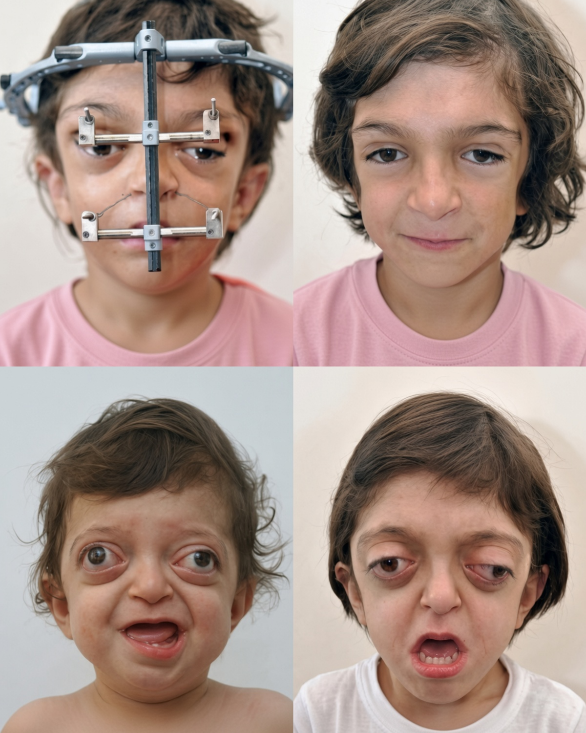
The four photographs circulating online show that sequence in one child. Early frames capture severe proptosis and midface hypoplasia. A later frame shows a rigid external distraction (RED) halo: a metal frame pinned to the skull and linked by rods and wires to the midface. The last frame shows a child whose eyes sit within protective rims, whose midface has come forward, and who can close the lids and smile without the earlier distortion.
That halo is not decoration. After a Le Fort III or monobloc osteotomy, surgeons do not yank the midface forward in one pull. They cut the bone, attach the RED, and turn activation screws a fraction of a millimetre a day. New bone fills the gap — distraction osteogenesis. Over weeks the orbits deepen, the airway lengthens, and the cheekbones move under the eyes so the globe is no longer hanging unprotected. Typical halo wear is on the order of seven to twelve weeks, sometimes longer, before the device is removed under a brief anaesthetic.
Treatment is staged across childhood, not a single operation. Infants may need fronto-orbital advancement or posterior vault expansion to give the brain room and drop intracranial pressure. Midface advancement usually waits until later childhood, when the child can tolerate the halo and when airway or eye protection becomes urgent. Many patients still need orthodontics and a Le Fort I or jaw-balancing procedure in the mid-teens once growth is nearly finished. Complications exist: pin-site problems, need to reposition the halo, incomplete correction, later relapse of midface position. Teams at high-volume craniofacial centres judge outcomes by airway, eye coverage, occlusion, and the child’s own comfort — not only by photographs.
The viral collage is a compressed version of years of planning, CT-guided cuts, nightly screw turns at home, and follow-up that lasts into adulthood. The calm smile at the end is real. So is the work that made room for the brain, covered the eyes, and let a face grow into something a child can live behind.
Sources
Boston Children’s Hospital and Seattle Children’s Hospital clinical overviews of Crouzon syndrome
The Craniofacial Center description of RED halo Le Fort III distraction
Journal literature on Le Fort III distraction osteogenesis with rigid external devices in Crouzon syndrome (including midface and airway outcomes)
GeneReviews / FGFR craniosynostosis syndromes overview
Medscape craniofacial syndromes imaging and surgical sequence notes