When the Nightmare Returned: Ka’Vayah’s Fight with a Rare Brain Vascular Malformation
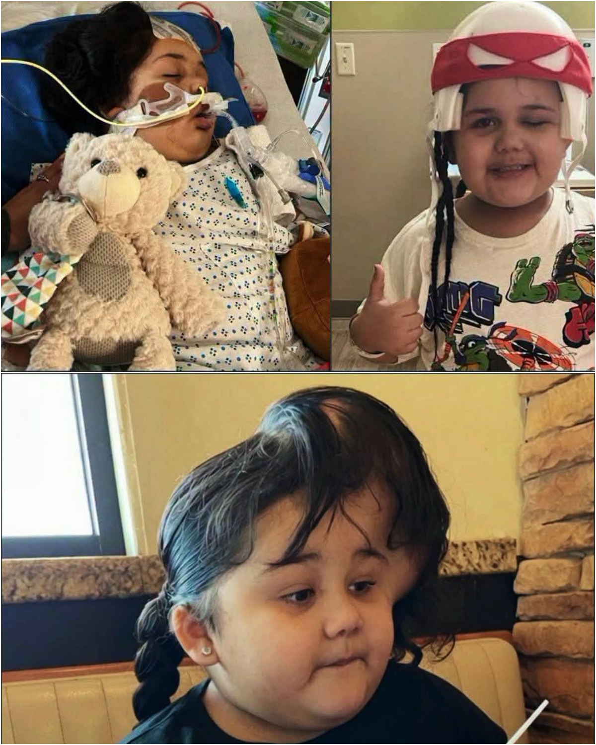
At four years old, Ka’Vayah’s childhood changed in an instant. Sudden seizures sent her family rushing for answers. Doctors discovered a rare tangle of abnormal blood vessels in her brain—an arteriovenous malformation or similar cavernous malformation—that had ruptured. What followed was urgent surgery, then a series of additional brain operations, including one that lasted 11 hours. Years of rehabilitation followed as she worked to regain strength and stability. Her family believed the worst chapter had closed.
A recent check-up delivered news no parent wants to hear: the condition had returned.
Pediatric brain vascular malformations such as AVMs and cavernomas are uncommon. They form when arteries and veins connect abnormally, creating fragile clusters that can bleed without warning. In children a rupture can trigger seizures, stroke-like symptoms, or life-threatening hemorrhage. Treatment often requires a combination of microsurgery to remove the lesion, sometimes preceded by embolization to reduce blood flow, and later reconstructive steps. Recovery can take years; protective helmets are sometimes needed while the skull heals or while residual risk remains. Recurrence or residual malformation, though not inevitable, is a recognized possibility that demands lifelong imaging follow-up.
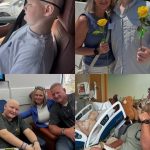
The photographs that accompany Ka’Vayah’s story capture both the ordinary joy of a little girl with braided hair and the medical reality of hospital beds, oxygen, and a protective helmet. They illustrate what families living with these diagnoses already know: progress is real, but it is rarely linear.
Public details about Ka’Vayah’s specific case remain limited to the account shared by her family. Broader medical literature documents similar pediatric journeys—emergency evacuation of hematoma, lengthy resections, infection risk, and the emotional weight of a “returned” finding on a routine scan. Outcomes vary widely depending on location, size, and how completely the malformation can be treated.
For families in this position, the immediate next steps typically include further imaging, multidisciplinary review (neurosurgery, interventional radiology, neurology), and planning that balances risk of another bleed against the risks of additional surgery. Support from pediatric rehabilitation teams and other families who have walked the same path often becomes as essential as the operating room itself.
Ka’Vayah’s story is still unfolding. It is a reminder that even after the longest operations and the hardest years of recovery, vigilance remains part of the care.
Sources
- Family account accompanying the photographs
- Medical literature and hospital patient stories on pediatric arteriovenous malformations and cavernous malformations (Alder Hey Children’s Hospital; Jackson Health System / Holtz Children’s; UCSF Benioff; Journal of Neurosurgery: Pediatrics)
- General references on presentation, rupture risk, and multimodal treatment of childhood brain vascular malformations