Pediatrics and Precision: The Complex Separation of Conjoined Twins Arthur and Heitor in Brazil
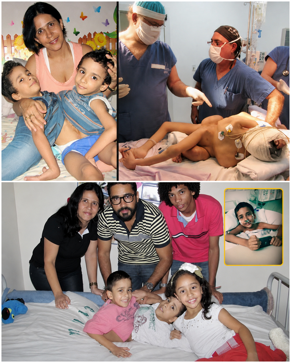
In April 2009, twins Arthur and Heitor Brandão were born at the Hospital Materno Infantil (HMI) in Goiânia, Brazil. The boys, whose family was originally from Riacho de Santana in Bahia, were conjoined from the thorax through the abdomen and pelvis (a complex thoraco-omphalo-ischiopagus configuration). They shared a liver and genitalia and had three legs between them, one of which was malformed. Their shared anatomy created significant circulatory and developmental challenges that required years of specialized care.
The family relocated to support intensive medical follow-up. Over roughly six years, the boys underwent multiple preparatory procedures—reports cite around 15 operations in total, including placement and management of tissue expanders. These silicone devices gradually stretched the skin to create enough coverage for reconstruction after separation, a process often compared to the progressive expansion seen in pregnancy. Nutritional support and careful monitoring were essential because the twins were initially too fragile for early separation, which is typically preferred in the first year of life when feasible.
On February 24–25, 2015, a large multidisciplinary team led by pediatric surgeon Zacharias Calil performed the separation at HMI. The operation lasted approximately 14–15 hours (with total anesthesia time reported around 18 hours in some accounts) and involved dozens of specialists—commonly cited figures range from more than 40 to 51 professionals, including surgeons, anesthesiologists, and support staff. Surgeons divided the shared liver, addressed the thoracic and pelvic connections, reconstructed abdominal walls, and used tissue from the third leg for repairs where possible. Advanced planning tools, including 3D prototyping of organs, aided the precision required for the intricate anatomy. During the procedure, the team discovered that each twin had a separate intestine, bladder, and gallbladder—findings that differed from some preoperative assumptions.
Both boys were transferred to intensive care in critical condition, requiring mechanical ventilation and showing high fever in the immediate postoperative period. Three days later, Arthur suffered cardiac arrests and died. Heitor remained in serious but improving condition; he eventually recovered, was discharged months later, and has continued under long-term follow-up. Heitor later returned for routine evaluations, and accounts from subsequent years describe him living with adaptive mobility support after losing one leg in the separation process.
The case highlighted both the extraordinary technical demands of separating complexly conjoined twins and the emotional and ethical weight carried by families and medical teams. Years of preparation, tissue expansion, imaging, and coordinated surgical expertise made the procedure possible, even as the outcome underscored the high risks involved when vital organs and vascular systems are shared. Heitor’s survival and ongoing life illustrate the potential for independence after such interventions, while the loss of Arthur remains a somber part of the story.
This Brazilian case stands as a notable example of pediatric surgical collaboration and the progressive use of preparatory techniques such as tissue expanders and three-dimensional planning in high-stakes conjoined-twin separations.
Sources
- G1 (Globo) reports on the twins’ birth, preparation, surgery, Arthur’s death, and Heitor’s recovery (2015–2016).
- Estadão and other Brazilian outlets covering the February 2015 operation and immediate postoperative course.
- Contemporary accounts detailing team size, duration, shared anatomy, tissue expanders, and surgeon Zacharias Calil’s role.