The Miracle of Jaxon Taylor: How Surgeons Reconnected a Toddler’s Skull After Complete Occipitocervical Dissociation
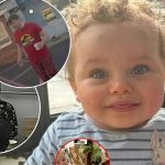
On 15 September 2015, Rylea Taylor was driving her two children through northern New South Wales when their car collided head-on with another vehicle at about 110 km/h (70 mph). She and her nine-year-old daughter Shayne survived with serious but treatable injuries. When she pulled 16-month-old Jaxon from the wreckage, she knew immediately that his neck was broken.
The crash had fractured his top two cervical vertebrae and torn the ligaments that hold the skull and upper spine together. His head and first vertebra were completely detached from the rest of the spinal column — a complete occipitocervical dissociation, popularly called “internal decapitation.” His spinal cord was bent at roughly 45 degrees and extremely vulnerable. Most patients with this injury die at the scene or never breathe or move again. Jaxon was still breathing on his own.
He was immobilized with extreme care and airlifted to Lady Cilento Children’s Hospital in Brisbane, where he came under the care of senior spinal surgeon Dr Geoff Askin. Imaging confirmed one of the worst injuries of its kind Askin had ever seen. Because Jaxon’s spinal cord had not been severed, there was a narrow window for reconstruction.
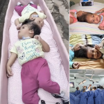
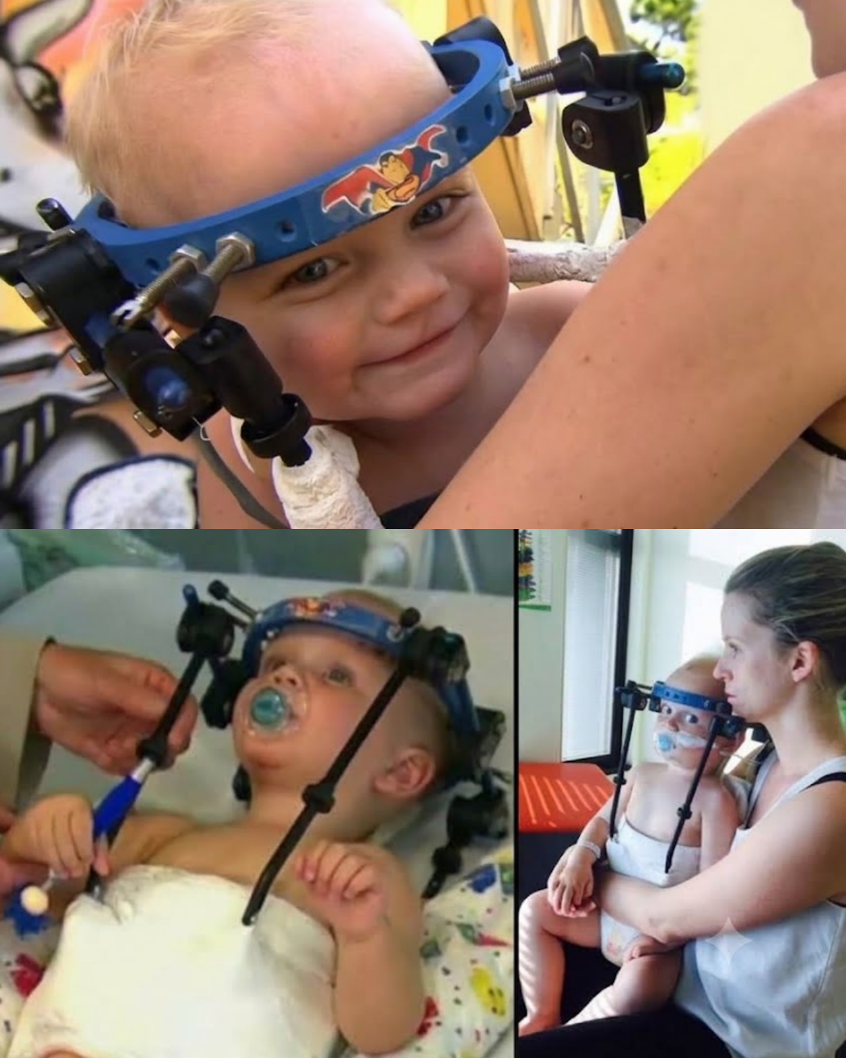
The six-hour operation began with a custom halo vest screwed into the toddler’s skull to lock his head and neck in position. Askin’s team then reduced the dislocation millimetre by millimetre under high-definition magnification. Standard screws were too large for Jaxon’s tiny bones, so surgeons used fine surgical wire to reconnect the base of the skull and the axis (C2). A small piece of one of Jaxon’s own ribs was harvested as a living bone graft to fuse the unstable segments. The rib would later regenerate.
Jaxon woke without paralysis. Within two to three weeks he was taking unsteady toddler steps, holding a parent’s finger. He wore the halo brace for about eight weeks to three months while the graft and wiring consolidated; local hospitals sent X-rays to Brisbane so Askin could confirm alignment. Doctors expected him to walk, run and live a normal life, with the main long-term caution being avoidance of high-impact sports that could cause another whiplash-type injury. Shayne also recovered after surgery for abdominal and spinal injuries.
Askin and the family called the outcome a miracle — not because the technique was unknown, but because the spinal cord had survived an injury that usually destroys it. Pediatric bone and neural tissue are more forgiving than adult tissue, yet the survival of both respiration and motor function after complete dissociation remains rare. A 2010 review of similar upper-cervical dislocations found that most victims die before diagnosis; of those who reach hospital, many never regain independent breathing. Jaxon was among the small minority who did.
The case drew global coverage because it combined extreme trauma, meticulous pediatric spinal surgery, and an almost complete functional recovery. It also underscored two practical lessons: the protective value of proper child restraints (Jaxon’s body stayed strapped while his head was flung forward) and the importance of absolute immobilization from the roadside onward.
Today the story is remembered as one of the most striking pediatric spinal reconstructions of the last decade: a 16-month-old whose skull was separated from his spine, rebuilt with wire and a sliver of his own rib, and who walked again.
Sources
Scientific American, “How Surgeons Reattached a Toddler’s Head” (K. J. Lee, 19 Oct 2015)
CBS News, “Skull and spine reconnected in 16-month-old Australian boy, Jaxon Taylor” (7 Oct 2015)
SBS News, “Toddler has head reattached to spine after ‘internal decapitation’” (8 Oct 2015)
7 News Queensland / Katrina Blowers reporting (Oct 2015)
Contemporary coverage in The Irish Times, UPI, CBC, NBC News, and Newser summarizing the same hospital and surgical details.